FIGURES
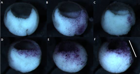
Figure 1: Preliminary trial of DN-XRhoA microinjection. Un-injected control (A); control embryo injected with only 200pg/blas. β-Gal mRNA (B); embryo injected with 184pg/blas. DN-Rho mRNA (C); embryo injected with 368pg/blas. DN-Rho RNA (D); embryo injected with 552pg/blas. DN-Rho RNA (E); embryo injected with 552pg/blas. DN-Rho RNA showing extensive cell death and necrosis (F, see along the white bar). All DN-Rho RNA-injected embryos were also injected with 200pg/blas. β-Gal mRNA.
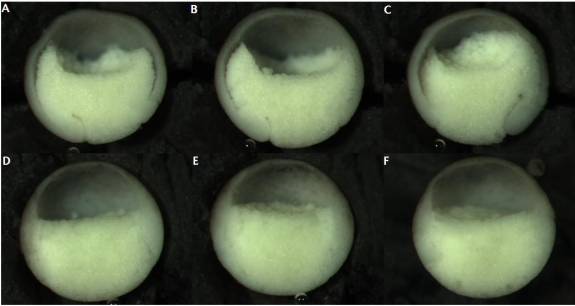
Figure 2: ROK-inhibited embryos allowed to recover in 0.1X MBS before fixation. Un-injected control embryos (A-C); embryos injected into the blastocoel with 23nl ROK inhibitor at stage 8 (D-F).
A

B
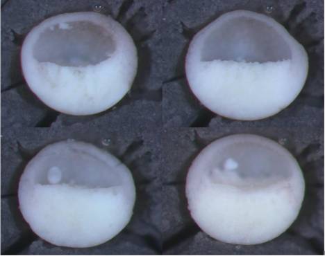
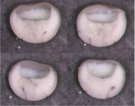
Figure 3: Normal (A) and un-rescued (B) controls from the DN-Rho-phenotype rescue experiment, using Active-V14 XRhoA. The un-rescued embryos were injected with 250pg/blastomere of DN-XRhoA N-19 RNA.
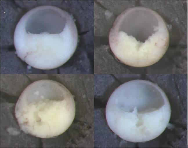
Figure 4: Rescued embryos from the experiment using Active-V14 XRhoA. These were co-injected with 18.4pg/Blasof Active-V14 XRhoA RNA along with the DN Rho RNA (250pg/Blas).
A
B
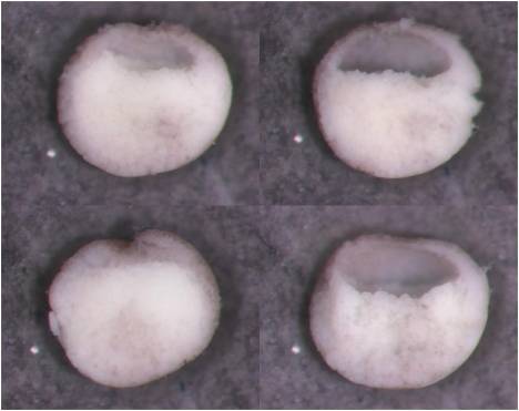
Figure 5: Normal (A) and un-rescued (B) controls from the DN-Rho-phenotype rescue experiment (1st attempt), using Wildtype XRhoA. The un-rescued embryos were injected with 250pg/blastomere of DN-XRhoA N-19 RNA.
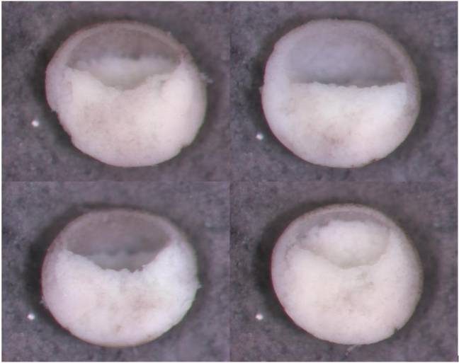
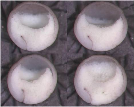
Figure 6: Rescued embryos from the experiment using Wildtype XRhoA (1st attempt). These were co-injected with 765pg/Blas of Wildtype XRhoA RNA along with the DN Rho RNA (250pg/Blas).
A
B
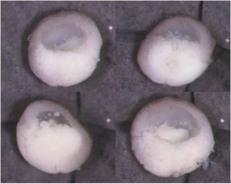
Figure 7: Normal (A) and un-rescued (B) controls from the DN-Rho-phenotype rescue experiment (2nd attempt), using Wildtype XRhoA. The un-rescued embryos were injected with 300pg/blastomere of DN-XRhoA N-19 RNA.

Figure 8: Rescued embryos from the experiment using Wildtype XRhoA (2nd attempt). These were co-injected with 918pg/Blas of Wildtype XRhoA RNA along with the DN Rho RNA (300pg/Blas).
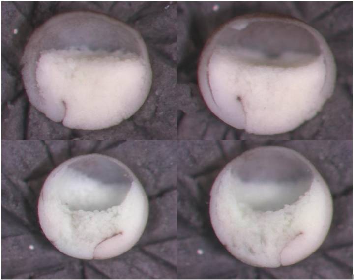
Figure 9A: Control embryos from the myosin inhibition assay using Blebbistatin. These embryos were kept in ficoll during the half-hour period it took to inject the embryos under experiment, as well as during the one-hour period, the experimental embryos were allowed to heal the needle punctures. All of the above steps were carried out at room temperature. The top two pictures are from a batch of embryos that showed less vegetal-cell-upwelling at stage 11 than normal.
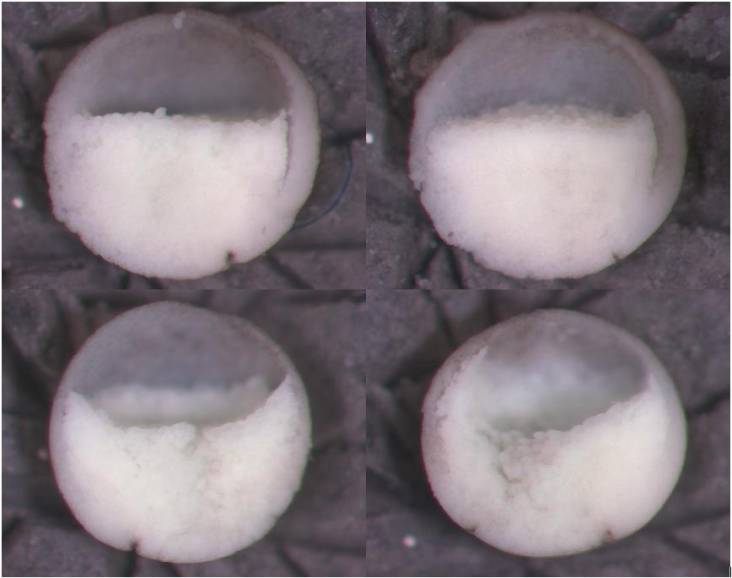
Figure 9B: Embryos injected with 23nl of 16.67mM Blebbistatin in the Blastocoel cavity at stage 9. Notice the stunted archenteron development compared to the controls (Figure 9A). The top two pictures are from a batch of embryos that showed less vegetal-cell-upwelling at stage 11 than normal.