RESULTS:
Rescuing Dominant Negative XRhoA Phenotype using Constitutively-active XRhoA
Constitutively-active RhoA (V14) was already known to be a very toxic molecule before we attempted to use it in rescuing the DN-Rho phenotype (Nagel, personal communication). During earlier rescue trials, all of the embryos injected with V14-RhoA RNA died out well before the desired stage (stage 11). Embryos were then injected with subsequently lower doses of V14-RhoA starting from ~180pg/blastomere, to as low as 18.4pg/blastomere, however, no significant rescue from the phenotype was observed. The embryos (n = 20), showed somewhat weaker phenotype as compared to the un-rescued controls, nonetheless there was a very wide range of appearances ranging from dead (1/4th) to odd shaped, mushy in texture, that crumbled easily during fracturing, despite having been fixed.
Rescuing Dominant Negative XRhoA Phenotype using Wildtype XRhoA
Wildtype XRhoA, when co-injected with DN-Rho into the dorsal blastomeres at 4-cell-stage, slightly weakened the phenotype, at concentrations three times more than those of DN-Rho. This change was more apparent when a higher than the ‘required minimum’ (last term’s work) amount of DN Rho was used; that is 300pg/Blas rather than 250pg/Blas, countered by a proportionately higher dose of wildtype Rho RNA. When more than the required minimum (250pg/Blas) of DN was used, although the rescued embryos looked no better than the previous trials, but the contrast between the rescued and the un-rescued embryos was more well defined.
In both sets of embryos (n1 = 35 & n2 = 25), while some showed undeniable signs of rescue, few still appeared inhibited and there were also those that could not be accurately classed as rescued on un-rescued because they looked somewhat in between the two, and still others bore chunks of dead tissue (see charts 1 & 2). The death rate was particularly high when 300pg of DN-XRhoA RNA was used. The features that might be associated with the rescue were thinning of the Blastocoel roof due to intercalation of cells, tissue separation at the Brachet’s cleft, and some faint upwelling of the vegetal cells. The embryos deemed ‘indistinct’ showed no more than one of these features but most such embryos had no significant tissue separation at Brachet’s cleft or any hint of vegetal cell upwelling. The archenteron however, in most rescued embryos was no more than a small notch. The thinning of the Blastocoel roof was especially more apparent when 300pg/Blas of DN RNA was used, countered by 918pg of Wildtype RNA. The higher level of death and necrosis in these trials might be the result of a large amount of exogenous RNA exhausting the cells metabolism, or could be due to some effect of the wildtype Rho RNA.
Chart 1
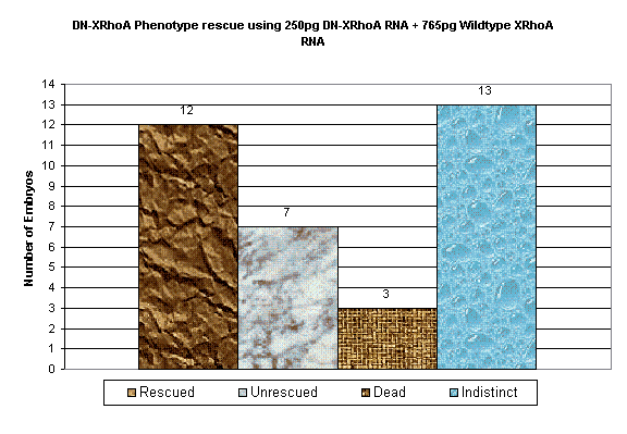
Chart 2

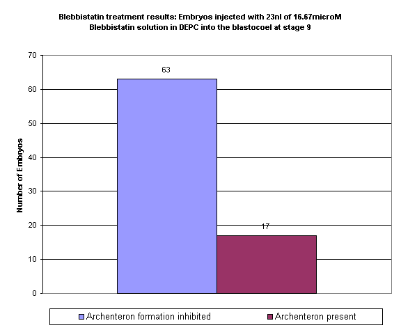
Effect of Blebbistatin on Vegetal Rotation:
Blebbistatin, a myosin inhibitor, when injected into the Blastocoel of stage 9 embryos, affected archenteron formation and hence involution of the marginal zone cells. Vegetal rotation, although somewhat weakened, still occurred in Blebbistatin treated embryos. The archenteron in most treated embryos was no more than a small notch (see chart 3).
Chart 3