|
 |
Bronchiectasis
Bronchiectasis is a fixed, irreversible dilatation of the bronchial tree. Morphologically it is classified as (1) tubular, (2) saccular/cystic or (3) fusiform.
Pathophysiology: infection (eg. tuberculosis), chronic fibrosis and traction, hereditary or congenital causes (eg. cystic fibrosis).
Predisposing co-factors are smoking, pollutants, pneumoconioses, muco-ciliary transport defects (eg. ciliary dyskinesia, Kartagener's syndrome), immunosuppression, chronic steroid therapy, chronic lung disease.
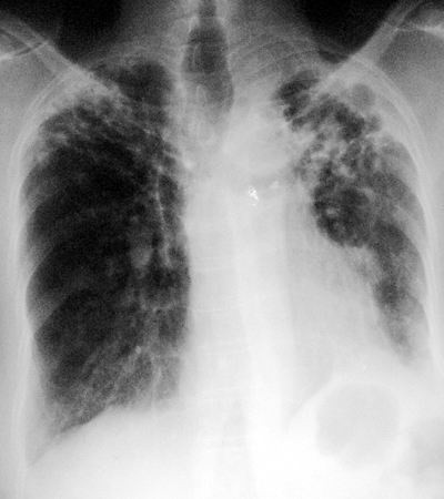
CXR Findings: dilated, often thick walled bronchi, in short axis presenting as multiple ring densities.
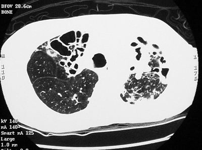
CT Findings:
- dilated, thick walled bronchi often with areas of traction and bronchial crowding
- "signet rings" - dilated thick walled bronchus (ring) with adjacent often dilated pulmonary artery (diamond)
Bronchiectasis may be fairly diffuse (eg. cystic fibrosis), or focal (middle lobe or lingula as in middle lobe syndrome).
“Aunt Sophies”: Usually fairly typical.
- Any small cystic or bullous lung process, regional or diffuse
- LAM, eosinophilic granuloma if there is a dominant cystic component
The problem is usually one of undercalling or not seeing the findings on the plain films. Do high resolution CT when in doubt.
|
