Sarcoidosis - Stage II
Pathophysiology: See Sarcoidosis Stage I.
A
progression of the lung disease including the presence of non-caseating
granulomata, interstitial lung changes along with adenopathy.
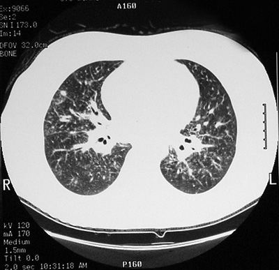
CXR/CT Findings:
-
small nodular
changes in the upper 2/3 of the lung, along with increased
linear interstitial lines
-
the nodular changes
may be endobronchial, peribronchial or perivascular giving a
beading effect, which is seen on high resolution CT
-
hilar and
mediastinal adenopathy: there may be less symmetry than on
earlier stage I disease
-
the changes have a
central perihilar predilection and spread peripherally
ōAunt Sophiesö: a gamut of mid and upper lung small
nodular/interstitial disease with adenopathy:
1.
metastatic lung disease
2.
inhalational lung disease: silicosis, berylliosis
3.
primary lung cancer, nodal metastases and central
adenopathy
4.
lymphoma (nodules usually larger and less in number)
5.
drugs: eg. methotrexate
6.
granulomatous infection: tuberculosis, fungal infections
7.
pulmonary amyloidosis, less common
|