|
This technique uses the digital capabilities along with the multiple tube
angulations available on some GI Fluoroscopic units (e.g. Phillips MD4) for
decubitus views. The filling phase of this examination has been discussed in
the other double contrast barium enema sections.
Equipment:
- 750 cc of Polibar 100%
- Rectal catheter with silicone balloon
- Air or carbon dioxide (infused using hand pump or pressure regulated
infusion pump)
|
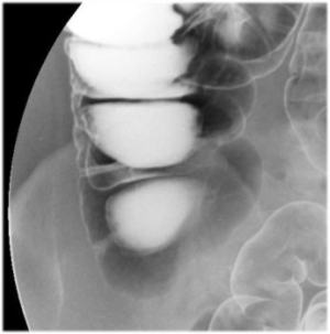
Upright Position |
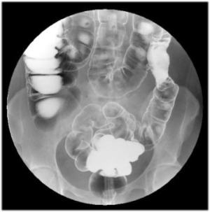
| Rectum (12"
FOV) |
 |
| Hepatic Flexure (12Ē
FOV) |
 |
| Splenic Flexure (12Ē
FOV) |
 |
|
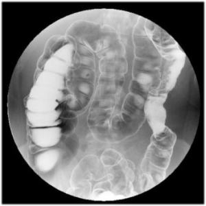
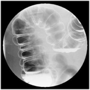
Right Lateral Decubitus
Position |
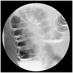
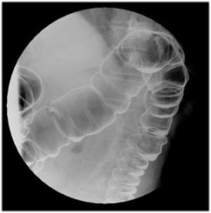
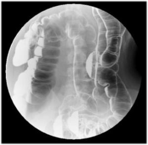
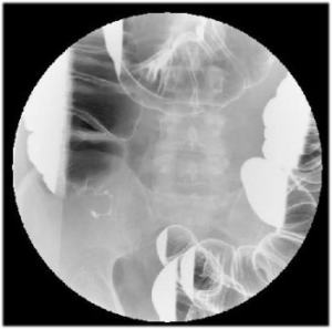
Survey
View of the Entire Colon:
- Survey view of entire colon
- Use 16Ē FOV
- Must include the entire colon
|
 |
|
 |
|
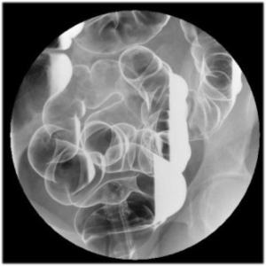
Left Lateral Decubitus
Position |
Survey
View of the Entire Colon:
- Survey view of entire colon
- Use 16Ē FOV
- Must include the entire colon
|
 |
|
 |
|
 |
|
Supine |
Survey
View of the Entire Colon:
- Survey view of entire colon
- Use 16Ē FOV
- Must include the entire colon
|
 |
|
 |
|
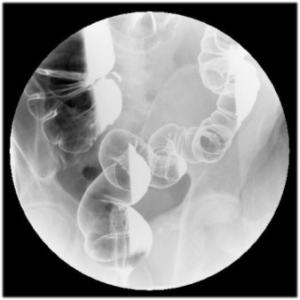
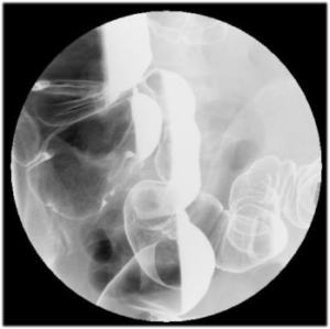
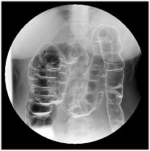
Sigmoid Views |
Sigmoid
Views:
- Angled views (cephalad and/or caudad views)
- Decubitus views (right and left)
- Use 12Ē FOV
|
 |
 |
 |
|
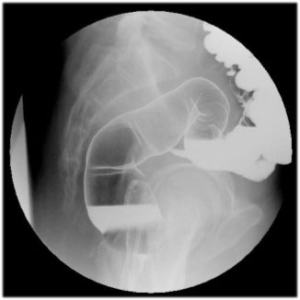
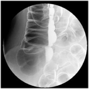
Rectum Supine Shoot Through Lateral View |
Rectum
Supine Shoot Through Lateral View:
- May deflate the rectal tube
- Do not forget to REINFLATE
the balloon
|
 |
|
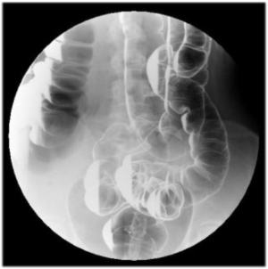
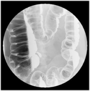
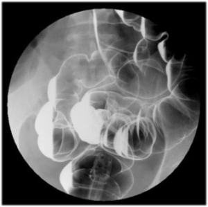
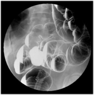
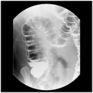
Cecum Views |
-
Use 12Ē and/or 9Ē FOV
-
May have to place patient on
Trendenlenburg position
-
Angle tube
-
Prone view may sometimes drain
barium out of the cecum
-
Views should include supine,
right and left decubitus
|
 |
 |
 |
|
 |
|
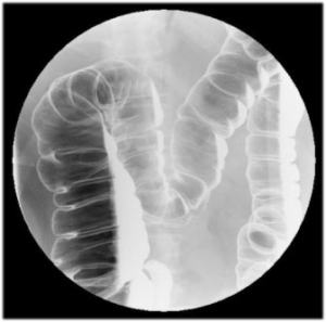
Upright Views |
- AP survey of entire colon
- Use 16Ē FOV
|
 |
|
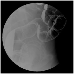
Upright Oblique Views of the Hepatic
Flexure |
- Hepatic flexure - turn patient to left (LPO)
- Use 16 & 12Ē FOV
|
 |
|
 |
|