Images:
Selected images are shown below. Click to
enlarge. |
 |
 |
|
Case
Summary
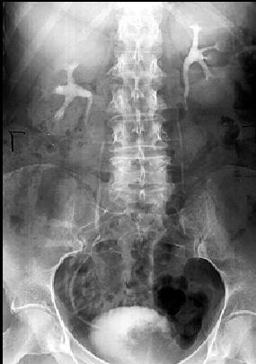
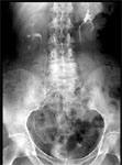
Diagnosis: Bladder Carcinoma
Clinical hints:
This patient was typical, presenting with hematuria.
He had a transitional cell carcinoma. Chronic cystitis and
stones, smoking, and rarely analine dye exposure, are risk
factors.
Radiological Pearls:
- typical filling defect on IVP
- irregular bladder mucosa
- U/S is best for screening
- CT and MRI best for staging
Films were kindly provided by Dr. G. Cooke.
|