Images:
Selected images are shown below. Click to
enlarge.
|
|
 |
|
|
|
 |
|
Case
Summary
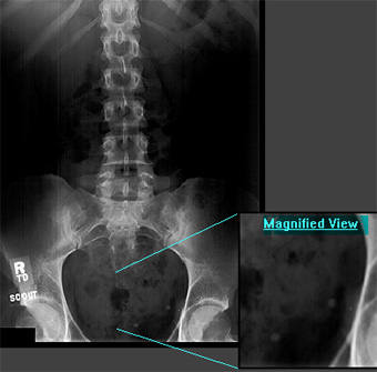
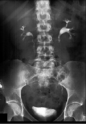
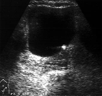
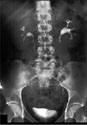
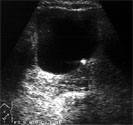
Diagnosis: Stone at the Left Ureterovesicular
Junction
Clinical hints:
This patient had a left kidney stone which lodged in the
ureterovesicular junction. Edema led to mild dilation of the
left collecting system. He passed the stone later on.
Radiological Pearls:
- 85% of renal stones calcified
- IVP & U/S: site & obstruction
- retrograde study: site & removal
- stones < 5mm: pass; > 10 mm: no
Films were kindly provided by Dr. R. W. McCallum.
|