Images:
Selected images are shown below. Click to
enlarge.
|
|
 |
|
|
|
 |
|
Case
Summary
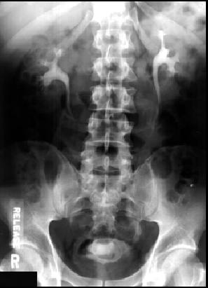
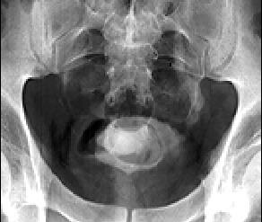
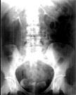
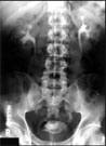
Diagnosis: Simple Ureterocele(s)
Clinical hints:
Seen in young patients most often as an incidental finding.
25% patients develop stones. May have a history of
microhematuria.
Radiological Pearls:
- typical "filling defect(s) in bladder
- "lucent line" around defect(s)
- may see stone in ureterocele
- may see dilated ureter
Films were kindly provided by Dr. R. W. McCallum.
|