|
Chest Tomogram
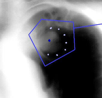
This view is a supine plain tomogram of the mass in the left
upper lobe. The mass itself (dark blue dot) is
surrounded by air (light blue dots) around almost its whole
circumference.
Decubitus views would have shown that this mass was mobile
within the cavitary space. |