Images:
Selected images are shown below. Click to
enlarge. |
 |
 |
|
Case
Summary
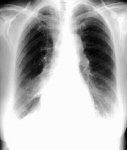
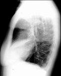
Diagnosis: Middle Lobe Pneumonia
Clinical hints:
Typical clinical presentation of pneumonia. When recurrent,
may indicate structural disease of the middle lobe bronchus,
or middle lobe bronchiectasis.
Radiological Pearls:
- consolidation of the middle lobe
- right heart border silhouetted out
- may have associated atelectasis
- bronchiectasis likely, if recurrent
Films were kindly provided by Dr. W. J. Weiser.
|

