Images:
Selected images are shown below. Click to
enlarge.
|
|

|
|
|
|
 |
|
Case
Summary
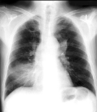
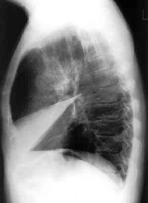
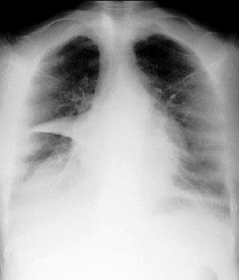
Diagnosis: Middle Lobe Pneumonia and Atelectasis
Clinical hints:
This patient had three lung diseases: acute middle lobe
pneumonia; tuberculosis in the right upper lobe (chronic); and
a carcinoma in the left supra-hilar area.
Radiological Pearls:
- often seen with atelectasis
- lordotic view very useful to assess
- may be seen with bronchiectasis
- if recurs, rule out central cancer
Films were kindly provided by Dr. W. J. Weiser.
|