Images:
Selected images are shown below. Click to
enlarge. |
 |
 |
|
Case
Summary
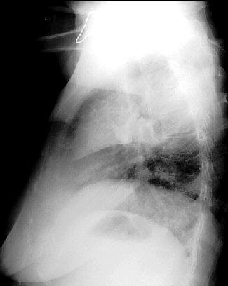
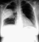
Diagnosis: Pneumococcal Pneumonia
Clinical hints:
Typical "lobar" consolidation is not commonly seen today. The
clinical picture is one of pneumonia, but the x-ray will
seldom indicate the specific etiologic agent.
Radiological Pearls:
- classic air-space consolidation
- lobar or segmental, classically
- usually non-specific changes
- effusion common
Films were kindly provided by Dr. W. J. Weiser.
|