Images:
Selected images are shown below. Click to
enlarge. |
|
 |
|
 |
 |
|
Case
Summary
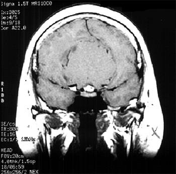
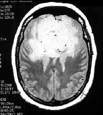
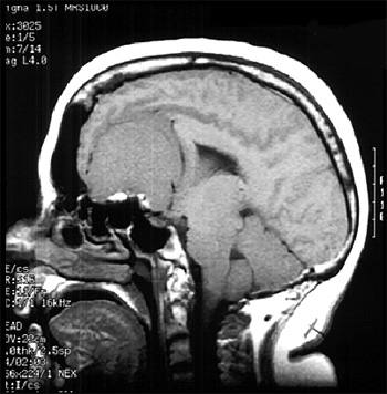
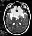
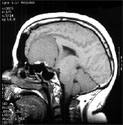
Diagnosis: Meningioma
Clinical hints:
This patient had a meningioma of the planum sphenoidale. A
common extra-axial tumor with typical features on imaging. The
tumor is surgically removed with a good prognosis.
Radiological Pearls:
- extra-axial tumor, typical sites
- calcification typical
- CT "screening" imaging
- MRI best imaging modality
Films were kindly provided by Dr. K. Terbroog.
|