Images:
Selected images are shown below. Click to
enlarge. |

|
|
 |
 |
|
Case
Summary
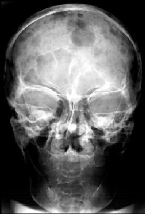
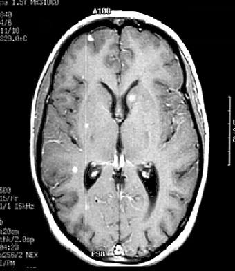
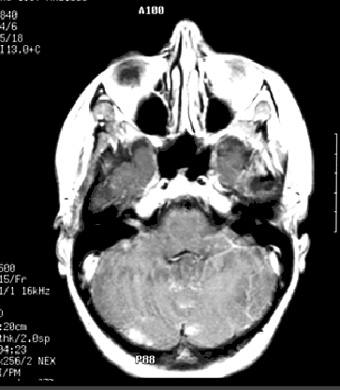
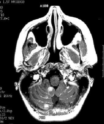
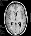
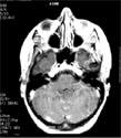
Diagnosis: Skull and Brain Metastases
Clinical hints:
This patient had a lumpectomy for breast carcinoma. The
imaging is typical. Metastases may occur many years after
treatment of the primary carcinoma.
Radiological Pearls:
- lesions usually lytic (85%)
- 15% breast ca. mets. blastic
- CT usually sufficient & diagnostic
- MRI best imaging modality
Films were kindly provided by Dr. K. Terbroog.
|