 |
|
Periodontium:
Anatomy and Histology Review
ALVEOLAR BONE
Portion of the alveolar process to which the PDL anchors the tooth (Note : Distinguish alveolar process from alveolar bone).
Alveolar process : Portion of the jaw bone which contains the teeth and the alveoli in which they are suspended. The alveolar process rests on basal bone. Development of the alveolar process is dependent on tooth eruption When teeth fail to develop (e.g. anodontia) the alveolar process fails to form. When all teeth are extracted most of the alveolar process becomes involuted.
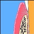
The alveolar process is composed of an outer cortical plate (compact bone). The alveolar bone lines the alveolus. It is a thin cortical plate with numerous perforations (cribriform plate) that allow the passage of blood vessels between the spongiosa and the PDL. The coronal rim of the alveolar bone forms the alveolar crest which generally parallels the CEJ at a distance of 1-2mm.
RADIOGRAPHIC FEATURES
Radiographically, the alveolar bone appears as a continuous, radiodense line separated from the tooth surface by a radiolucent periodontal ligament space. The radiographic equivalent of the alveolar bone is the lamina dura. Where roots lie close to the outer cortical plate, the cortical plate and the alveolar bone may fuse, thereby eliminating the intervening cancellous bone. Occasionally the thin bone overlying a root may resorb, thereby producing a fenestration of a dehiscence.
Maxillary posterior teeth may extend close to the maxillary sinuses and even extend into the sinus, with only a paper-thin lamella of bone separating the PDL from the respiratory mucosa lining the sinus.
For details on the microscopic features of the cells and matrix of compact an cancellous bone, review lecture on “Bone”.
CLINICAL CONSIDERATIONS
Through remodeling, the alveolar bone may become displaced in relation to the alveolar process, thereby allowing tooth movement to take place.
Interruptions in the continuity of the lamina dura in the apical region of an alveolus are of diagnostic significance in the identification of periapical lesions.
Proximity of the alveolar bone to sinus cavities or major nerves (mandibular nerve) may create problems during tooth extraction or surgical interventions.
Following tooth extraction, the alveolar bone tends to resorb, a process that may compromise the placement of endosseous dental implants and affect the construction of removable prostheses.