Convergent extension is the most important morphogenetic movement involved in the construction of the primary body axis during early embryogenesis. Convergent extension is a key event both during gastrulation, when convergent extension of the marginal zone occurs to create the anterior-posterior axis, and during neurulation, when convergent extension of the central region of the neural plate, occurs as the neuraxis elongates and the neural tube closes. During convergent extension the embryo extends along a single anterior-posterior axis through forceful intercalation of the cells of the epithelium, which results in the embryo to become a football shape from originally nearly spherical shape.
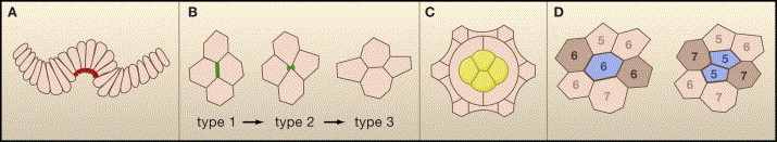
The intercalation is the major driving mechanism of convergent extension. Cells in an epithelium are able to move within the plane of the sheet, thereby changing their neighbour relationships without violating the integrity of the epithelium. . The intercalating cells extend basal protrusions which squeeze between their opposing neighbors beneath their adherens junctions. As the intercalating cells move forward, these protruding tips become broader in the anterior¨Cposterior and dorsoventral dimensions, effectively ¡°plowing through¡± the adherens junctions and forcing an opening for the remainder of the intercalating cell to insert between the contralateral cells. Fig 1 and Fig 2 show schematic pictures and experimental photos of epithelial cell intercalation.
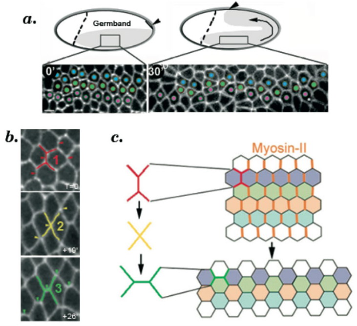
Myosin-dependent
junction remodeling
controls planar cell intercalation and axis elongation.
Bertet. C., Sulak. L., and Lecuit. T. Nature 429:667-671(2004)
¡¡
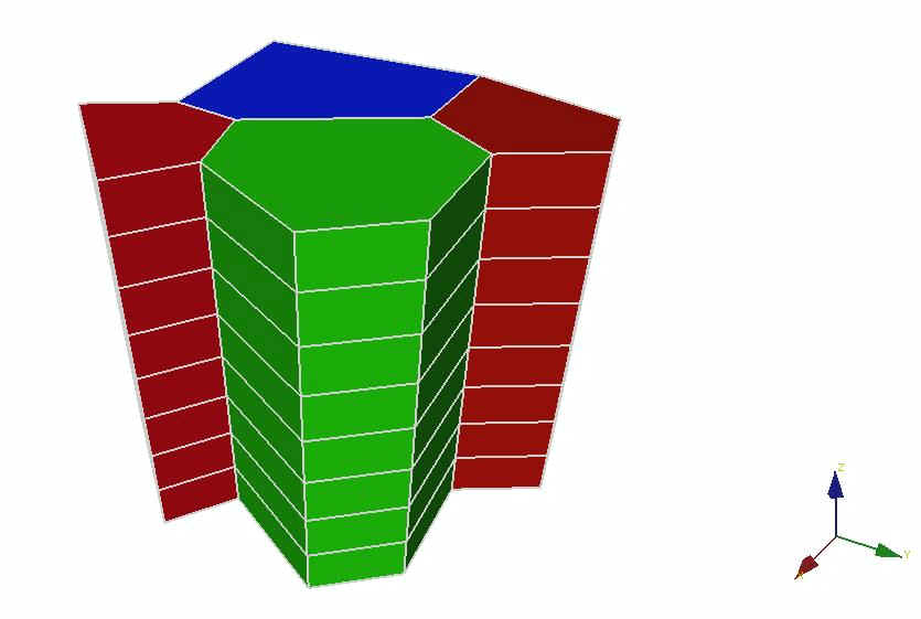
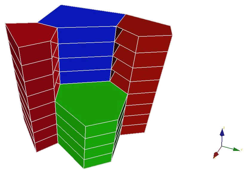
A custom 3-D finite element analysis package SIMBA was used and numerical simulation was carried out at Engineering Biomechanics Group at University of Waterloo during summer 2006. Four columnar epithelial cells were modeled and the driving force for the intercalation was assumed to be the surface tension acting on the peripheral surface (except for apical and basal surface) of the columnar cells. The difference of surface tension between different cells will drive them to change neighbours.