|
|
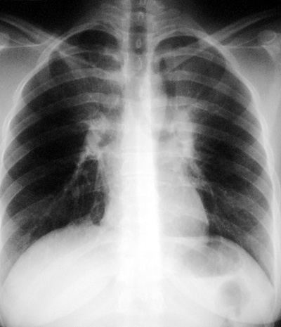
Left Upper Lobe Atelectasis
Common Pathophysiology:
- endobronchial cancer, rarely a metastasis
- endobronchial mucus plug (eg asthmatics, bronchopulmonary aspergillosis)
- constriction of bronchus by tumor (small cell, lymphoma), bronchostenosis
- endobronchial tuberculosis or other granulomatous infections (fungus)
CXR Findings: Can be subtle and easy to miss especially when only partial atelectasis is present. Typically, presents as an ill-defined haziness/density in left upper lung with obscuration of the aortic arch.
Other findings on chest x-ray include elevation of the left mainstem bronchus, variable elevation of the left hemidiaphragm and typical retrosternal line seen on lateral view.
This radiograph is actually of a patient with bilateral upper lobe atelectasis.
Clues:
Look at both PA and lateral views
Look for ancillary findings (eg. adenopathy, TB, underlying airways disease)
Follow chronology of findings if old films available
"Aunt Sophies": Many especially if atelectasis partial
- Pneumonia
- Lung tumor
- Granulomatous infections eg tuberculosis
- Lung contusion
- Rarer entities: COP, eosinophilic lung disease, pulmonary emboli
|
