Mediastinal Lymphoma
Pathology: One of the more common causes of mediastinal adenopathy.
Imaging Clues: Bulky asymmetric adenopathy may be non–Hodgkin's while symmetric adenopathy may be Hodgkin's lymphoma. Need clinical and tissue diagnosis. Lung nodules, pleural effusions and pericardial effusions often seen in non Hodgkin's lymphoma.
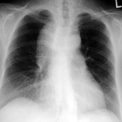
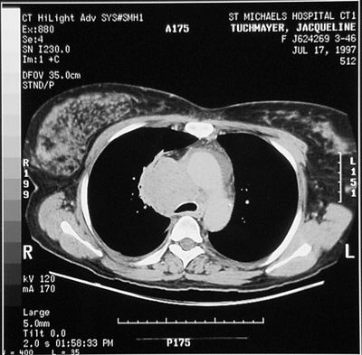
CXR/CT Findings:
- hilar and/or mediastinal adenopathy
- lung nodules or masses
- the larger masses may have typical air bronchograms within them
- pleural effusions
- pericardial effusions
Post Treatment Imaging:
Chemotherapy or radiation will kill the tumor cells and result in dystrophic calcification with the “sterile” node. Variable calcification with nodal masses may result.
Rarely, a treated nodal group may be firm enough because of fibrosis and/or calcifications that it causes atelectasis by impinging on bronchial walls. Atelectasis is quite uncommon in untreated lymphoma other than in end-stage presentations.
|