|
|
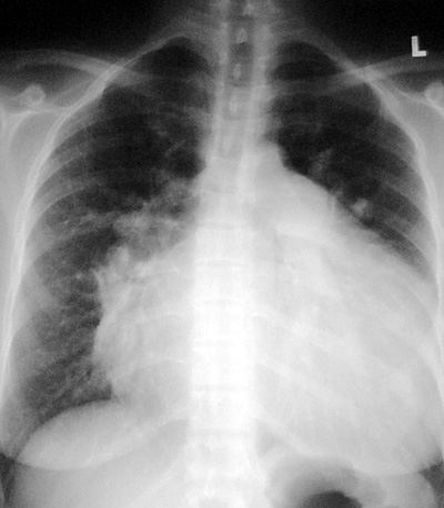
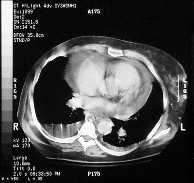
Pericardial Effusion
Pathophysiology: Usually an exudate, but transudative
effusions can occur.
Transudative effusions: CHF, renal failure, and
hypoproteinemic states.
Exudative effusions: Post-myocardial infarction, viral
pericarditis (Coxackie B), other infections especially
tuberculosis, collagen vascular diseases (SLE), post trauma (hemopericardium),
tumor (eg. lymphoma or metastases to pericardium), bleeding
diatheses and coagulopathies with hemopericardium.
CXR Findings:
-
Pericardial fat
stripe sign (classic sign): On lateral film: fluid within
pericardial sac (white) outlined on either side by pericardial
and endocardial fat (black). This results in a white central
stripe outlined by black bands seen along anterior of the
right ventricle.
-
enlarged
pericardial silhouette
-
may see pericardial
calcifications (eg. markers of old infection etcģ)
-
globular or
flask-like cardiac silhouette in cases of large effusions
Clues:
-
small effusions not
seen on CXR will be seen on CT
-
classic
ōpericardial fat stripeö, rarely seen
-
often the
differential diagnosis include causes of an enlarged cardiac
silhouette (see ōAunt Sophiesö)
-
echocardiography to
confirm/quantitate extent of effusion
ōAunt Sophiesö:
-
Cardiomyopathies
-
Multiple valve
disease, usually due to rheumatic fever, with multiple chamber
cardiac enlargement
-
Pericardial cyst,
metastases, lymphoma
|
