|
|
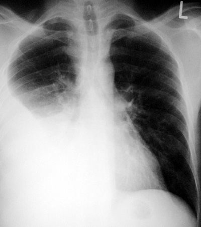
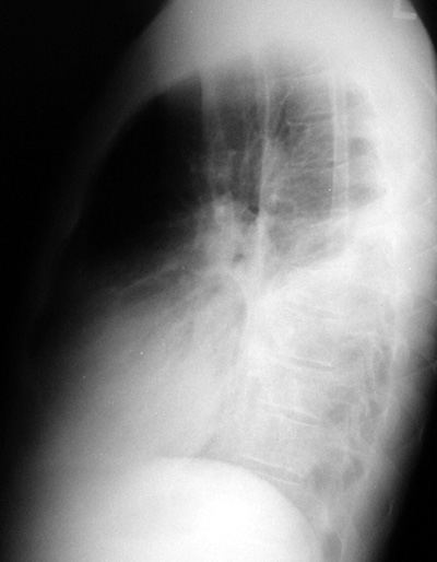
Pleural Effusion
Pathophysiology:
Transudative: Essential consists of water and electrolytes,
seen in CHF, renal failure, hypoproteinemic states and fluid
overload states.
Exudative: Multiple causes: infections (para-pneumonic
effusions with community acquired pneumonia CAP),
tuberculous effusions, hemothorax (bleeding of any cause),
malignant effusions (eg. metastases, lymphoma), pulmonary
embolism, collagen vascular diseases (SLE, rheumatoid lung
disease, scleroderma, mixed CTD).
Chylothorax: Lymph in the pleural space. Rarely seen.
Injury or disruption of the thoracic duct as seen in
lymphangioleiomyomatosis (LAM).
CXR Findings:
-
Classic “meniscus
sign” with pleural effusion curving to chest wall, seen on PA
and lateral films
-
Blunting of the
costo-phrenic sulcus
-
Hemithorax
“white-out”
-
Associated passive
or relaxation atelectasis in lung bases
-
May see mediastinal
shift in large effusions
Imaging Clues:
-
Need about 150 cc
of fluid in the pleural space to see on routine PA and lateral
films
-
Decubitus views may
show as little as 15 cc of fluid (rarely done as not suspected
on routine CXR)
-
Uncomplicated
effusions always associated with relaxation atelectasis. The
volume loss is more or less the same as the pleural effusion
in small to moderate effusions so overall no volume change in
hemithorax. In large effusions, the fluid volume is
larger than the atelectatic volume loss and mediastinum may
shift to contralateral side.
-
Big Clue: if the pleura is encased, fibrotic or thickened, or if the
mediastinum is involved with pathology. There will be
variations and exceptions to these principles.
-
Pseudo-tumor:
the effusion may encyst or loculate, simulating a lung mass or
tumor
“Aunt Sophies”:
-
Atelectasis
-
Pleural fibrosis
-
Pleural tumor (eg.
mesothelioma, metastastic disease)
-
Extrapleural fat
|
