Staphylococcal Pneumonia
Pathophysiololgy: Caused by staphyloccus aureus.
Usually spread by droplet inhalation or direct hematogenous
spread to lungs due to bacteremia or septicemia. Often
underlying staphylococcal infections or abscesses may be present
(e.g. kidneys, skin, bone).
A
suppurative exudative infection develops in lungs with
desquamated epithelial cells, leukocytes, and debris filling the
alveoli and airspaces. May also involve the airways leading to
bronchitis and bronchiectasis, and the pleura (parapneumonic
effusions). The pneumonia may undergo cavitation due to airways
communication. Rarely osteomyelitis may develop in the thoracic
cage.
Clinical Clues: Look for underlying staph infections in
skin, kidneys, bones, sinuses etc… Consider other causes of
cavitary lung disease. Need bacteriologic identification of
organism for definitive diagnosis.
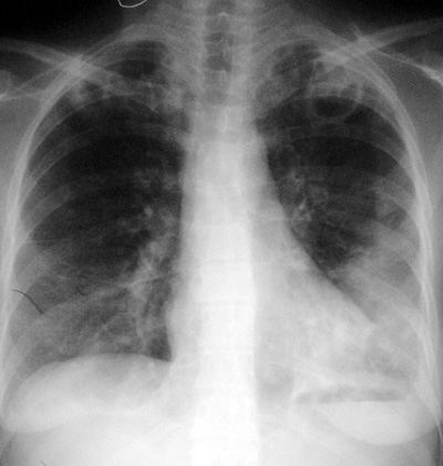
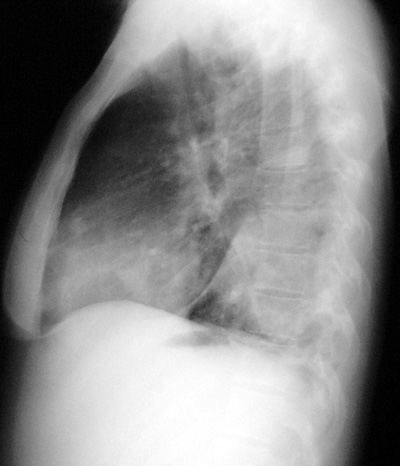
CXR Findings:
-
regional airspace
process (often with air bronchograms), non specific finding
-
cavitary areas,
often multiple with ill-defined walls and surrounding air
space disease (non–specific but suggestive)
-
pleural effusions
-
adenopathy NOT seen
-
very rarely, lytic
bone lesions (ostemyelitis, bone abscess)
Radiologic Clues: in clinical setting of a CAP, small
multiple cavitary areas should suggest staphylococcal pneumonia.
“Aunt Sophies”:
-
Gamut of focal air
space diseases (eg. infection, hemorrhage, tumor, aspiration,
etc…)
-
Gamut of cavitary
lung diseases
−
infections: TB, granulomatous infections, CAP etc…
−
tumor: squamous cell metastases
−
vasculitis: Wegener’s granulomatosis
−
trauma: traumatic pneumatoceles
−
complicated bullous disease: infected bulla
−
others: infected bronchogenic cyst(s)
|