|
|
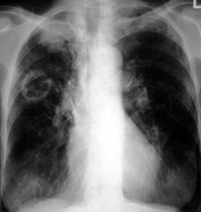
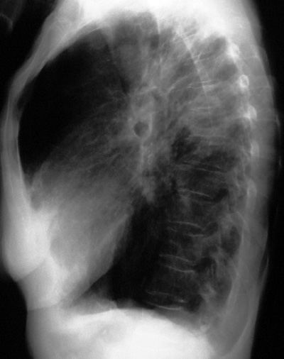
Tuberculosis - Reactivation
Pathophysiology: See Tuberculosis – Primary
Post primary tuberculosis is now felt to be largely reactivation
of dormant foci of tubercle bacilli in the lungs or elsewhere.
True re-infection tuberculosis is far less common.
Occurs in situations of host susceptibility: genetic or
in various host suppressed states: e.g. bad health habits
(indigents), high risk geographic areas (far East, Africa, India
etc…), immuno-compromised hosts (HIV), drug treatments
(steroids, chemotherapy).
The
dormant TB foci are most prevalent in the upper apices of the
lungs (especially the apical and posterior segment) and during
reactivation, an exudative hyperimmune pneumonia occurs. This
is characterized by tissue destruction, cavitation, airways
involvement with bronchiectasis, and marked fibrosis including
the pleural surfaces.
Clinical and Radiologic Clues: Think tuberculosis!
Commonest cause of failure to diagnose is the physician not
thinking of tuberculosis!
Tuberculosis, especially post primary TB, is a great imposter
mimicking many lung disease processes. Tuberculosis and
sarcoidosis are classic “Aunt Sophies”.
CXR/CT Findings:
-
upper lung, apical,
superior segment of lower lobe airspace disease; cavitary
-
calcified
granulomata (small nodules)
-
calcified hilar
lymph nodes
-
Ghon complex
-
Ranke complex
-
disease with
fibrotic changes and architectural distortion
-
pleural fibrosis,
hilar traction, traction of vessels and airways
-
pleural effusion
-
fibrothorax:
any cause
-
bronchiectasis:
upper lung, superior segment predilection
-
rarely may see
regional adenopathy in re-activation TB
-
lung nodules and
masses: may be non-specific but in upper lungs
-
pneumothorax,
broncho-pleural fistula with pneumothorax
-
empyema: encysted
pleural infection with Slit Pea Sign on CT
-
“tree and
budding” on CT
-
skeletal
tuberculosis: Pott’s disease, thoracic spine osteomyelitis,
paralumbar masses
-
rarely: rib
destruction and empyema leading to empyema necessitatis
“Aunt Sophies”:
-
granulomatous
infections: fungal, atypical tuberculosis
-
granulomatous
diseases: e.g. sarcoidosis stages III and IV; eosinophilic
lung disease; end stage hypersensitivity alveolitis,
silicosis
-
CAP: e.g. severe
gram negative, mixed aerobic and anaerobic pneumonias and lung
abscesses, Legionella pneumonia
-
COLD: complicated
emphysema, bullous disease, bronchiectasis of any cause,
cystic fibrosis
-
cavitary lung
disease gamut: infections, tumours, vasculitis, pneumatoceles
etc…
-
pleural effusions
and fibrosis: infections, tumours, asbestos-related pleural
disease and mesothelioma, hemothorax, causes of fibrothorax
-
miliary pattern (micronodular):
fungal diseases, mycoplasma pneumonia, sarcoidosis,
micronodular metastases (e.g. thyroid)
|
