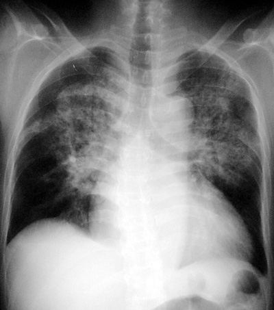
Pulmonary Edema - Alveolar
Pathophysiology: Airspace disease: any process which
replaces air in alveoli. The abnormal material may be: water,
blood, tumor, pus, aspirant, or surfactant. In cardiogenic and
non-cardiogenic edema, water, electrolytes, and proteinaceous
material fill the alveoli.
Clues: One airspace disease looks like any another and
differentiating them generally needs clinical correlation. Some
radiologic hints indicating edema are:
1.
perihilar or batwing symmetry of edema
2.
typical pericardiac and peridiaphragmatic lucent margins
(cardiac/renal edema)
3.
enlarged heart and valvular calcifications
4.
chronology: airspace disease which appears rapidly
(minutes) and clears rapidly (hours to days)
ôAunt Sophiesö: Any airspace disease
1.
Infections: all the CAP
2.
Tumor: bronchoalveolar cell cancer, lymphoma, metastases
3.
Bleeding: vasculitis (eg. Goodpastureĺs syndrome),
coagulopathies, trauma, anticogulants
4.
Aspiration pneumonia and chronic lipid pneumonia
5.
Acute silico-proteinosis, alveolar proteinosis
|