|
|
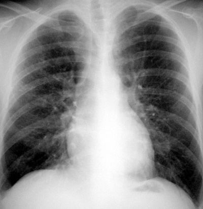
Pulmonary Histiocytosis X
Pathophysiology: An eosinophilic, non-caseating,
granulomatous, non-infectious inflammatory process of unknown
cause. This disease affects multiple organ systems including
the lung, bone, and central nervous system. The lesions are
granulomata and fibrosis in the lungs, destructive lytic lesions
in the skeleton, and inflammatory changes in the central nervous
system (pituitary or hypothalamus).
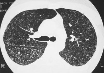
The
lung inflammation involves small centrolobular airways likely
with small airway involvement, nodular granulomata, air
trapping, cyst formation, and fibrosis.
Clinical Clues: Eosinophilic lung disease is generally a
disease of young male smokers. It should be considered
when a disease process is multisystemic and involves the lungs.
CXR/CT Findings:
-
Small nodular
mid-to-upper lung pattern
-
Associated with
small cystic areas in mid and upper lungs.
-
May improve, stay
stable, or worsen with fibrosis and end-stage lung findings.
-
Not associated with
significant adenopathy, pleural reaction, or airspace disease.
-
Classic High
Resolution CT findings: diffuse small centrolobular nodular
pattern and small diffuse cystic areas with a mid to upper
lung distribution.
-
Lytic lesions in
the thoracic cage (relatively unusual finding)
ôAunt Sophiesö: small nodular lung diseases with cystic
formation
-
Sarcoidosis (the
Ĺsyphilisĺ of the chest)
-
LAM (females, total
lung involvement, no discrete nodules)
-
Metastases (cystic
changes not dominant)
-
Small airways
bronchiectasis
-
Cystic fibrosis
Hint: Young smoking male, think eosinophilic lung
disease.
|
