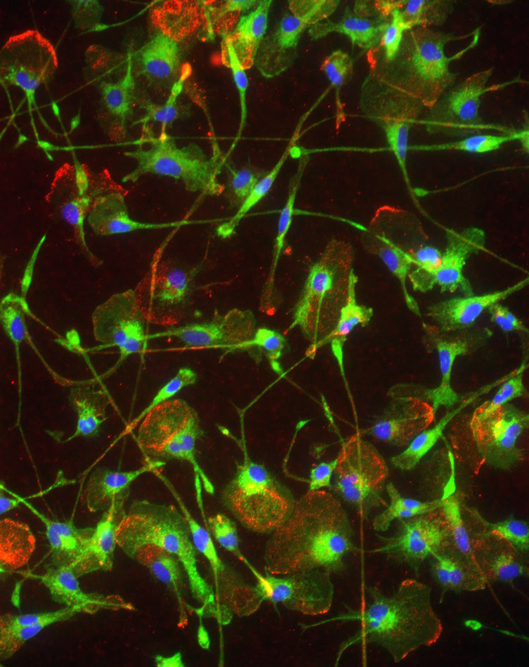
Microglia (the brain’s immune cell) evolved for nurturing, developmental and protective functions, but we now understand that they can contribute to brain damage. Microglial activation can be a ‘double-edged sword’; e.g., some growth factors they produce help during the repair phase after brain trauma, but they can also produce cytotoxic molecules that can reduce survival of damaged brain cells. Our in vitro studies focus on mechanisms of microglial activation, physiological and molecular outcomes, and roles of specific ion channels in these outcomes. The central hypothesis is that different ion channels contribute to specific aspects of microglial activation, and offer new therapeutic targets for controlling brain inflammation. Our published and ongoing studies support this hypothesis. For instance:
In our earliest studies, we discovered the first link between ion channels and microglial activation (Schlichter et al., 1996), and then developed ex vivo hippocampal ‘tissue-prints’ and which we showed that voltage-gated K+ channels (Kv1.3, Kv1.5) regulate microglia proliferation (Kotecha & Schlichter, 1999).
Later, we provided the first evidence that Kv1.3 and Ca2+/calmodulin-gated K+ channels in microglia regulate production of reactive oxygen species (Khanna et al., 2001). In addition to ion channels, we discovered that the Na+/Ca2+ exchanger reverses direction, promotes Ca2+ entry and activates production of reactive oxygen species [read article]; a mechanism that is well designed for microglial activation following an ischemic stroke.
We recently developed a powerful new in vitro model of the stroke penumbra, and used it to elucidate mechanisms and signaling pathways whereby ischemic neurons activate microglia, which then kill naďve neurons [read article].
We have found that Ca2+-gated K+ channels (SK3, SK4) regulate microglial activation through p38 MAP kinase. [read articles: here and here] SK channel blockers reduce their ability to produce harmful superoxide, nitric oxide and peroxynitrite molecules, and to induce apoptosis in bystander neurons. We have successfully translated results with an SK4 blocker (TRAM-34) from in vitro ? in vivo studies. Because an important function of microglia is to phagocytose foreign objects and cellular debris, a key discovery is that their molecular activation pattern during bacterial phagocytosis differs from, and modifies, the microglial response to other activating stimuli [read article].
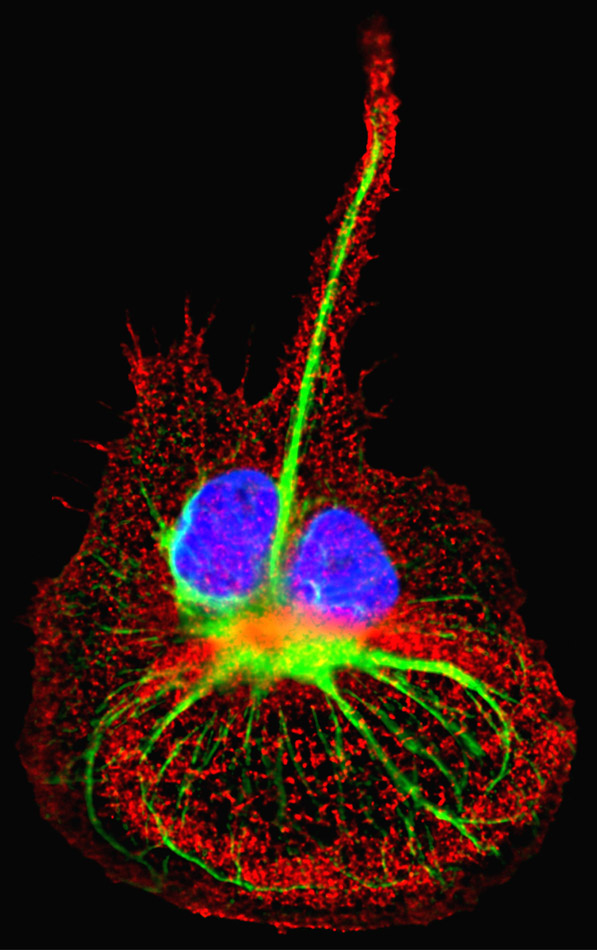
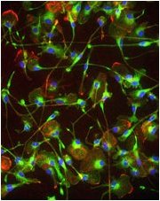
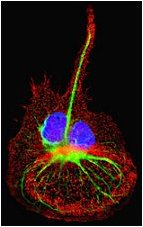
[Click the images to view them enlarged with descriptions.]
<-- Back