Approaches
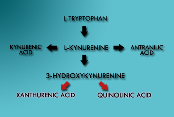
Diagram: Kynurenine pathway. In HD patients, there is a build up of 3-Hydroxykynurenine, which contributes to the pathophysiology of glial cells. Could this be a potential therapeutic target?
Protein Interaction Approach
There lies a kyurenine monooxygenase pathway in microglial cells that has identified to be a potential therapeutic target, as 3-hydroxykyurenine (3-HK), is increased in HD mice (27), proposing an alteration in the KMO pathway, which reduces enzyme activity and substrate affinity, attributing to the pathophysiology of glial cells (27). Hence, interruption of this pathway, or novel approaches in attempting to cease this prolonged excitotoxicity, may be of benefit to HD patients (28).
An unchartered approach...
NF-κB kinase complex activation is upregulated in HD mouse, a protein kinase pathway that’s a major integrator of cell signaling and response to cytokines (25), where mutant htt interacts with the IκB kinase complex, a key regulator of NF-κB (25). The number of pathways that can be influenced represents an interesting target for polyglutamine induced toxicity, with no evident approaches have been made in the field.
Immunological Approach
Examination of the peripheral immune system exhibited innate immune activation (22). Increased secretions of the ligand Interleukin-6 (IL6), is found to be increased in HD, whose secretion originates from monocytes or lymphocytes (25). Therefore, it is not entirely known whether degeneration begins in the PNS or CNS (25). Microglial cells, our nervous system macrophages, has increased activation as the disease progresses, and mutant htt is expressed in these cells (25), as interleukin-6 is a biomarker released. This fact was confirmed with flow cytometry and RT-PCR, in postmortem HD striatal tissue, confirming presence of IL-6, IL-8, and TNF-α, which alludes to an innate versus adaptive immune response (26), therefore, the immune system is a possible pathogenic pathway.
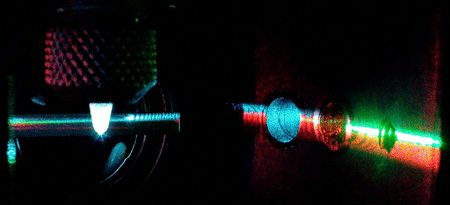
Photo: Flow cytometry can be utilized to assess cellular properties of HD cells, based on measures of fluorescence.
(Credit: University of Toronto, Immunology)