

Publications:
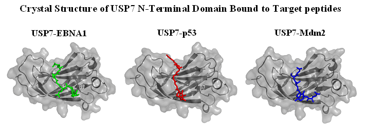
Sheng, Y., Saridakis, V., Sarkari, F., Duan, S., Wu, T., Arrowsmith, C.H. and Frappier, L. 2006 Molecular recognition of p53 and Mdm2 by USP7/HAUSP. Nature Structural and Molecular Biology. 13, 285-291. )
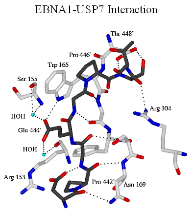
Saridakis, V., Sheng, Y., Sarkari, F., Holowaty, M.N., Shire, K., Nguyen, T., Zhang, R.G., Liao, J., Lee, W., Edwards, A.M., Arrowsmith, C.H. and Frappier, L. 2005 Structure of the p53-binding domain of HAUSP/USP7 bound to Epstein-Barr Nuclear Antigen 1: Implications for EBV-mediate immortalization. Mol. Cell 18, 25-36.
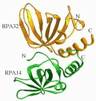
Bochkarev, A., Bochkareva, E., Frappier, L. and Edwards, A.M. 1999 The crystal structure of the complex of replication protein A subunits RPA32 and RPA14 reveals a mechanism for single-stranded DNA binding. EMBO J. 18, 4498-4504.
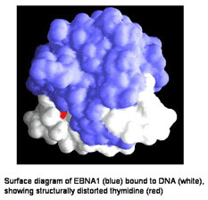
Bochkarev, A, Bochkareva, E., Frappier, L. and Edwards, A.M. 1998 2.2 Å Structure of a permanganate-sensitive DNA site bound by the Epstein-Barr virus origin binding protein, EBNA1. J. Mol.. Biol. 284, 1273-1278.
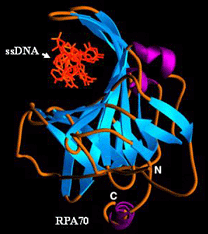
Bochkarev, A., Pfuetzner, R., Edwards, A. and Frappier, L. 1997. Crystal structure of the single-stranded DNA binding domain of replication protein A bound to DNA. Nature 385, 176-181.
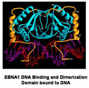
Bochkarev, A., Barwell, J., Pfuetzner, R., Bochkareva, E., Frappier, L. and Edwards, A. (1996) Crystal structure of the DNA binding domain of the Epstein-Barr virus origin binding protein, EBNA1, bound to DNA. Cell 84, 791-800.
Bochkarev, A., Barwell, J., Pfuetzner, R., Furey, W., Edwards, A. and Frappier, L. (1995) Crystal structure of the DNA binding domain of the Epstein-Barr virus origin binding protein, EBNA1. Cell 83, 39-46.