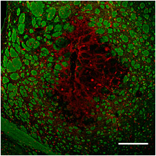
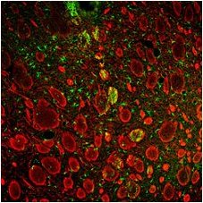
Inflammation and pathology after ischemic or hemorrhagic stroke.
Current work builds on our recently published
in vivo studies of ischemic stroke and intracerebral hemorrhage (ICH) in rats:
(2010) Evolution of Inflammation and White Matter Injury in a Model of Transient Focal Ischemia [read article]
(2010) White matter injury in young and aged rats after intracerebral hemorrhage [read article]
(2008) Glial responses, neuron death and lesion resolution after intracerebral hemorrhage in young vs. aged rats [read article]
(2008) Minocycline protects the blood–brain barrier and reduces edema following intracerebral hemorrhage in the rat [read article]
(2007) Evolution of the inflammatory response in the brain following intracerebral hemorrhage and effects of delayed minocycline treatment [read article]
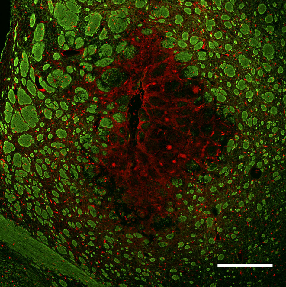
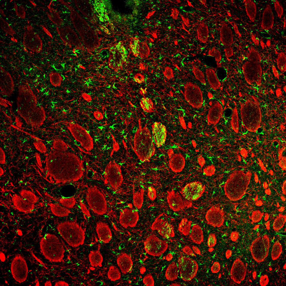
The central hypothesis is that brain inflammation is key to the delayed damage to neurons and white matter that follows a stroke. There is a clear correlation between inflammation and secondary damage after ICH, but cause-effect relationships have not been proven, nor are the individual contributions known for ‘innate’ immune cells: neutrophils, macrophages, microglia. Our current work focuses on the temporal and spatial development and roles of inflammation in damage to neurons and to white matter. We are comparing young and aged animals, with particular attention to early responses that include activation of microglia and infiltration of neutrophils and macrophages. By comparing ICH and transient focal ischemia in the same brain region (striatum), we have discovered differences in the evolution of damage and inflammation in the two forms of stroke.
There are several ongoing projects:
(i) effects of depleting blood neutrophils on blood brain barrier breakdown and white matter damage after a hemorrhagic stroke;
(ii) the first analysis of SC1, an extracellular matrix molecule involved in tissue remodeling, after ischemic and hemorrhagic strokes;
(iii) high throughput molecular analysis (microarrays, nanostrings, laser capture) to analyze inflammation and damage after ischemic and hemorrhagic strokes in old versus young animals.
[Click the images to view them enlarged with descriptions.]
<-- Back