 |
Samuel Richard Ph.D. student Medical Biophysics University of Toronto |
||
|
Research
Academic
Pretty fun
stuff
Coffee break stuff
|
Why use dual-energy x-ray imaging ?
The most important factor affecting a radiologist's ability to detect a
lesion in a radiograph (i.e., x-ray image) is not quantum noise
(caused by random photon fluctuations) but overlying anatomical structures such as overlapping
ribs and arteries. Computed tomography (CT) is an excellent way to
see through this clutter, however it is more expensive and causes much
more radiation to the patient (e.g., 1 chest CT exam is equivalent to ~300
radiographic chest exam). For this reason it important to search for simpler
and more cost-effective alternatives that can also provide
increased image quality. Dual-energy x-ray imaging significantly improves on
traditional radiography by being able to decompose a radiograph into different materials types,
thereby reducing the greatest factor affecting lesion conspicuity caused
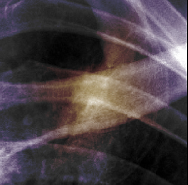
by anatomical clutter and overlapping structures. 1) A radiograph compresses all of the information unto a 2D plane.
2) A CT decomposes an image spatially along the horizontal (axial) dimension.
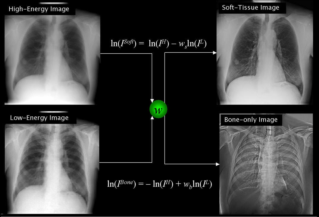
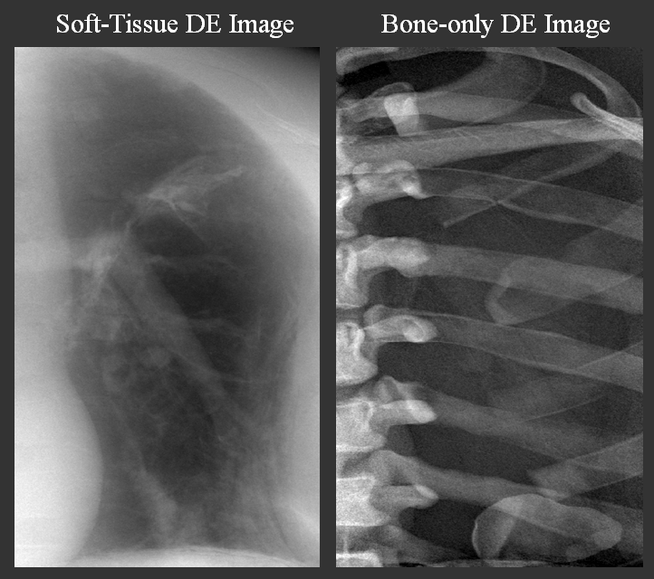
3) Whereas a DE image decomposes a radiograph into its components. Specifically, in the case of a chest radiograph the image is decomposed into a " soft-tissue" and "bone-only" DE image.
The physics of dual-energy imaging
Dual-energy imaging is a technique in which two
images acquired at different energies are processed to reconstruct images in
which bone or soft tissue are selectively canceled. It exploits differences in
the probability of photoelectric and Compton interactions in the object as a
function of x-ray energy and atomic number, with the photoelectric
cross-section exhibiting a stronger energy and Z dependence. Therefore, an
image acquired at lower energy (e.g., 60 kVp) will have higher bone contrast
than an image acquired at a higher energy (e.g., 120 kVp) due to calcium in the
bone.  A common algorithm for DE image reconstruction, derived from straightforward manipulation of Beer’s law, is weighted log subtraction. It can be shown that a soft-tissue-only image can be generated from a low- and high-energy image as shown above where Soft, H, L denote the “soft-tissue” dual-energy, high-energy, and low-energy images, respectively. The tissue cancellation parameter, w is a freely variable factor that weights the contribution of the high- and low-energy images in the reconstruction. A similar expression is written for the “bone-only” dual-energy image. References:
|
||


